Today, we know that treating cancer is not just about removing a tumor. It is also essential to help the immune system recognize and attack it more effectively. A research team led by Savieay Esparza and collaborators, published in Annals of Biomedical Engineering, studied a technique called high-frequency irreversible electroporation (H-FIRE) and discovered that it can do more than just destroy tumor cells: it can remodel the environment around the tumor and trigger signals that strengthen the body’s natural defenses.
H-FIRE therapy works by applying ultra-fast electrical pulses that perforate the membranes of cancer cells, causing their death, but without damaging other critical structures like nerves, blood vessels, or healthy tissues. In this study, the researchers went a step further: instead of aiming to eliminate the entire tumor, they applied a sub-ablative therapy, meaning they destroyed only part of the tumor (about 23%) to observe what changes would occur.
The treatment was tested in a mouse model of breast cancer. After applying the therapy, the scientists closely monitored what happened in the tumor, in the surrounding tissues, and in the lymph nodes that drain the tumor area over the following 1, 3, and 7 days.
The results were highly revealing. As expected, the tumor initially decreased in size. However, around the seventh day, it began to grow again. Although this could seem like a setback, it actually uncovered something much more important: the environment around the tumor had changed significantly.
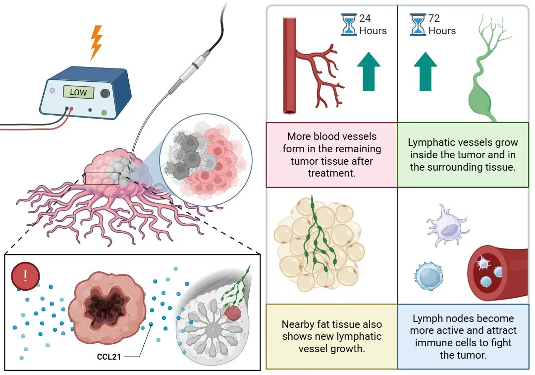
One day after therapy, there was an increase in the number of blood vessels within the surviving part of the tumor, suggesting that the tissue was trying to adapt or repair itself. Three days later, there was a noticeable increase in the number of lymphatic vessels, both inside the tumor and in the surrounding fatty tissue. Lymphatic vessels play a key role because they help drain fluids and guide immune cells where they are needed.
What was particularly striking is that these changes were not due to the usual growth factors like VEGFA or VEGFC, which are typically responsible for the formation of new blood and lymphatic vessels. Instead, the researchers detected a significant rise in a molecule called CCL21, a signal that acts like a “help call,” attracting immune cells such as dendritic cells and T lymphocytes to the area.

Figure 1. Sub-ablative electroporation promotes early blood vessel formation and subsequent lymphatic vessel expansion, fostering immune activation within the tumor microenvironment.
Moreover, the lymph nodes that drain the tumor area also showed changes. Although some differences were subtle, the blood and lymphatic vessels in these lymph nodes became more complex, and there was less collagen, which could make it easier for immune cells to move within them. The levels of CCL21 were also higher, while VEGFC decreased, reflecting similar patterns seen within the tumor.
These findings suggest that SA-HFIRE therapy not only partially damages the tumor but also prepares the surrounding environment for a stronger immune response. The therapy seems to set off changes that are not based on the typical vessel growth factors, but rather on pathways that activate and recruit the immune system.
The work of Savieay Esparza and colleagues opens new possibilities for the future, where combining H-FIRE with immunotherapies could lead to better outcomes in cancer treatment. The key would be to take advantage of the moment when the body is most ready to recognize and attack the tumor with greater strength.
Main Reference:
Esparza, S., Jacobs, E., Hammel, J. H., Michelhaugh, S. K., Alinezhadbalalami, N., Nagai-Singer, M., Imran, K. M., Davalos, R. V., Allen, I. C., Verbridge, S. S., & Munson, J. M. (2025). Transient Lymphatic Remodeling Follows Sub-Ablative High-Frequency Irreversible Electroporation Therapy in a 4T1 Murine Model. Annals of biomedical engineering, 53(5), 1148–1164. https://doi.org/10.1007/s10439-024-03674-y
Other References:
Cannon, R., Ellis, S., Hayes, D., Narayanan, G., & Martin, R. C., 2nd (2013). Safety and early efficacy of irreversible electroporation for hepatic tumors in proximity to vital structures. Journal of surgical oncology, 107(5), 544–549. https://doi.org/10.1002/jso.23280
Rossmeisl, J. H., Jr, Garcia, P. A., Pancotto, T. E., Robertson, J. L., Henao-Guerrero, N., Neal, R. E., 2nd, Ellis, T. L., & Davalos, R. V. (2015). Safety and feasibility of the NanoKnife system for irreversible electroporation ablative treatment of canine spontaneous intracranial gliomas. Journal of neurosurgery, 123(4), 1008–1025. https://doi.org/10.3171/2014.12.JNS141768
Li, S., Chen, F., Shen, L., Zeng, Q., & Wu, P. (2016). Percutaneous irreversible electroporation for breast tissue and breast cancer: safety, feasibility, skin effects and radiologic-pathologic correlation in an animal study. Journal of translational medicine, 14(1), 238. https://doi.org/10.1186/s12967-016-0993-7


